The equestrian community for horse people everywhere
Hoof Structure
The amount of information available about horse hooves and their trimming, shoeing, barefooting, wrapping, booting, soaking, and “orthotic-ing” is mind-boggling. So the first thing I want you to know in this post is that I’m not going to summarize all that or wade in to the “natural hoof care” debate or encourage you to purchase a product of some sort. My goal in this post is the same as it is for the blog as a whole: to offer you a different point of view on a subject — one that doesn’t seem to be readily available otherwise — that is based in biomechanics and good, solid biology. It will be your job to take what you learn here and figure out how you want to integrate it with what you already do.
This post will discuss specific aspects of the biology of hoof structure, and then the next post will consider functional aspects of those structures in terms of basic biomechanics. In both posts, the place we’re heading is a consideration of the nature and size of the element called the “coffin bone” by horsepeople and the “terminal phalange” by anatomists, and how that element functions in the stress environment of a large animal’s hoof.
We’ll start by looking at what bones are in a horse’s legs and feet. These bones have names as well as “definitions” based on where they’re located in the body and what other bones they connect to on the ends. For instance, animals have a lower jaw bone called the mandible. In mammals, the mandible is entirely mde of a bone called the dentary, which bears teeth on its upper surface (if the animal has teeth in its lower jaw at all, which some do not). There is a right dentary bone and a left dentary bone, one on each side of the animal’s lower jaw, and they join each other in the middle at the front (to form what we would consider the animal’s “chin”). The two halves are “sutured” there (that is the anatomical term) by a joint that is fixed or immovable and that looks like a wavy line of back-and-forthing such as might be done by a needle and thread. As the animal ages, this line disappears and there seems to be only one bone instead of two. On the back end of each mammal’s dentary bone there is a process that sticks upward, that makes a fairly high crest and that has a lower piece sticking back with a roller joint on it. When the lower jaw is put in proper life position against the skull, this roller joint articulates or fits against a socket in the skull, and the jaw rolls to open or close the mouth at this joint. The high crest projects up through a part of the skull that sticks out from the rest of the bone as a big arch. In life, it can be seen that strong jaw muscles attach to both the crest of the dentary bone and that arch on the skull. These muscles help to pull the jaws together so the teeth can chew food. Here is what the dentary looks like in a horse, when it is not attached to the skull.
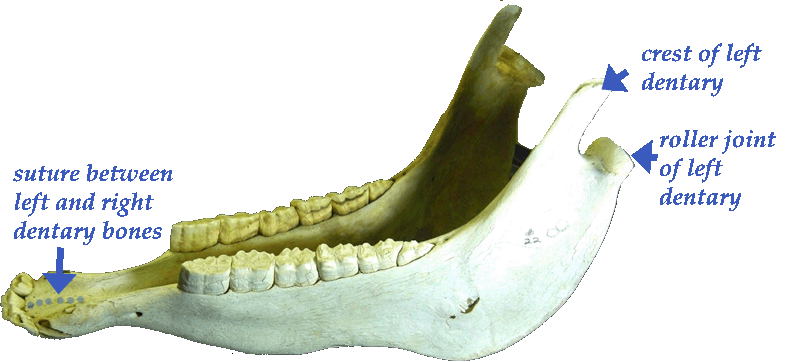 The picture below (left) shows you how the dentary bones look in position, articulated with the skull. You can see the crest goes through an arch on the skull and that the roller joint articulates with or fits up against a part of the skull that is shaped to receive it. The dentary in this picture is the white (uncolored) bone. The lower teeth are hidden by the upper teeth, which “lap” around them to the outside as this is drawn.

The skull to the right of the horse, above, is that of a human. It shows the human dentary bone and the way it fits to its skull, for comparison. I have colored the dentary a light bluish-purple. The point is to notice that the basic shape, position, and relationship to the bones around it are the same for the human dentary bone as in the horse. That’s why we consider both bones to be dentary bones, is that they meet the same “criteria” of definition for that bone. The dentary of the horse is therefore said to be homologous to the dentary of humans. It should be added that homologous structures may be seen to develop from the same embryonic tissues during development of different animals. So the similarity of structure that we call homologous reflects a real commonality of genetic origin and development.
Comparative anatomy gives us powerful tools for understanding animal structures. That’s probably more true for the hoof than for just about any other structure, because we all know how vitally important a horse’s hoof is. “No hoof, no horse” is an adage that’s been around about as long as people have been riding and working horses. And it turns out that the hoof is an astonishingly “little” bone.
Here is a horse skeleton with the bones of its front leg colored in a specific way. The circle I’ve drawn is around the bone horsepeople call the cannon bone, to serve as a place-marker with which you’re familiar. I’m not going to worry here about what other names horsepeople have coined for different parts of a horse’s limb anatomy, because that will actually confuse matters. I’m going to use terms that tell us about the homologies of these structures instead. Next to the horse skeleton is the skeleton of a human arm. I have colored the bones there so they correlate to their homologues in the horse skeleton. As we run through these bones, you can use your own arm and shoulder to help you learn the structures experientially.

At the top of the of the arm, the first good-sized bone that moves the arm in a mammal, is the scapula or shoulder blade bone. It’s colored pale turquoise here. In a horse, the scapula lies beneath (and gives the sloping ridge-shape to) the withers. In a human, the scapula is “down behind” our arm in our back because of the way our chest is flattened out side to side. There is a bone called the clavicle or collar bone in humans that is shown in white on the diagram above, that is not present in the horse. Then the next main bone in the sequence is the humerus bone. We looked at the humerus bone already in our discussion of the trotting gait in horses, and it’s colored in red on the diagram. It’s also in red on the human skeleton. In you, the humerus is inside your upper arm. (You’ll have to disregard the horse’s back leg here, on which I previously colored the femur red as well for a previous blog. The horse’s back leg is homologous to a human’s leg, too, but we are going to focus on the foreleg and arm as our example for this blog.)
Beneath (or “outward from”) the humerus there are two bones, side by side: the ulna and the radius. Here, I have colored the ulna a light green and the radius yellow. In humans (and many other mammals), the two bones are totally separated their entire length. They twist back and forth across one another when we rotate our forearms to turn our palms up or palms down. (If you sit with your right arm bent at the elbow, with your right hand just above your lap, you can move your arm in a way that allows you to feel these two bones. Put the fingers of your left hand across your right arm, about halfway between the inside of your elbow and your wrist. Press down firmly. Then, without moving your upper arm, simply twist your wrist to rotate the palm of your right hand so it faces the floor, then faces the ceiling again. (Your right thumb will face left when your hand is palm-down, and right when your palm is face-up.) The fingers of your left hand should feel the radius cross over the top of the ulna when you do this. The ulna is the longer of the two bones, and the back end of it “sticks out” to form our elbow.
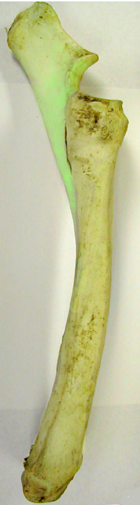 Horses have an ulna and radius, too, but theirs are fused together not too far below the elbow area. (The next time you saddle your horse, take a good look at the part of the horse’s arm just in front of the cinch or girth. You will see a very obvious elbow there.) It’s hard to see what they look like in the skeleton above, so here’s a closer picture of just those two bones in a horse. I colored the ulna light green and the radius yellow. You can see the dark line between them near the top of the radius, and then you can see that the ulna fuses to, and essentially becomes one with, the radius farther down the shaft. This is because the separate ulna and radius allow the hand to be twisted around the long axis of the leg — as is so in us — and that would be a really bad way for a horse’s leg to move. Can you imagine what would happen if a horse was running at 30 miles an hour and suddenly twisted its front hoof on its leg? So there is an adaptation of this structure that keeps the twisting motion from taking place. The radius and ulna are “locked” down.
Beneath the radius and ulna are the wrist bones, or carpals. In the big skeleton, those are not even drawn in, they are of such little consequence to many horsepeople learning anatomy. But of course, those bones are extremely important and you sometimes hear about one or more of them being injured in a performance horse. Since we’re heading for the hoof, though, we’re going to skip the carpals for now and head on downward. So the carpals are simply white on the human skeleton as well.
Beneath the carpals are the finger bones. The first of these is a long metacarpal bone that is actually positioned inside the fleshy part of the hand (where the palm is). Then, at the end of each metacarpal, there is a series of three little bones, each called a phalange. There are three phalanges on each finger except the thumb, which has only two phalange bones. If you take a look at your hand, you will see all this. On the human skeleton, I have colored the metacarpal a dark blue, the first phalange dark red, the second dark green, and the last orange or dark gold. However, I have only colored the bones of the middle finger — because that is the finger homologous to the hoof bones of a horse.
Look at the colors of the bones in the skeleton above, showing the horse hoof. You will see that the cannon bone is actually a metacarpal. And the bones that make up the pastern and the hoof are phalanges. Here is a diagram that shows you the end of this sequence a little more clearly. It’s a photograph of a horse hoof that was injected with plasticine so it could be sectioned cleanly and show you the structures. On the left is an image without any modification, and on the right is the same image with the bones colored so they correspond to the key I’ve just run through. (The diagram, from Wikimedia commons, is labelled in German.)

What should blow you completely out of any sense of complacency about your horse’s abilities, here, is realizing how very tiny the bone is that’s inside your horse’s hoof. I mean, LOOK at it! 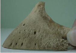 Here’s a nice shot of this coffin bone from a horse care website, that happens to have the edge of a person’s thumb and thumbnail in it, for scale. Take a look at just how small this bone really is. And if you consider the fact that the “hoof bone” or “coffin bone” is, in fact, a terminal phalange — the last tiny bone of a single finger — the fact that it bears so much weight, running at high speeds is nothing short of astonishing.
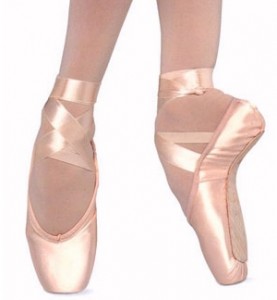
We are impressed, and rightfully so, by ballet dancers who leap and pivot en pointe, with their feet positioned so that their weight is borne only on the tips of the toes. Depending on the gender of the dancer, they may weight perhaps 80 to 150 pounds a person. What you can see here is that horses are also en pointe, minus the ballet slippers, but carrying closer to 1000 or 1200 pounds when they run, jump, and spin on those toe tips. It takes nothing away from human ballerinas, but I think it certainly adds to our appreciation of horses.
Or at least, I hope it will after we consider the consequences this has for the stresses (force per unit area) in the bones of horses’ feet. Coming up, next time . . .
Dawn Adams, Ph.D.
Copyright 2012 Dawn Adams, Ph.D. Do not reproduce in any format without written permission from Dr. Adams.
Views: 1912
Comments are closed for this blog post
The Rider Marketplace
International Horse News
© 2026 Created by Barnmice Admin.
Powered by
![]()